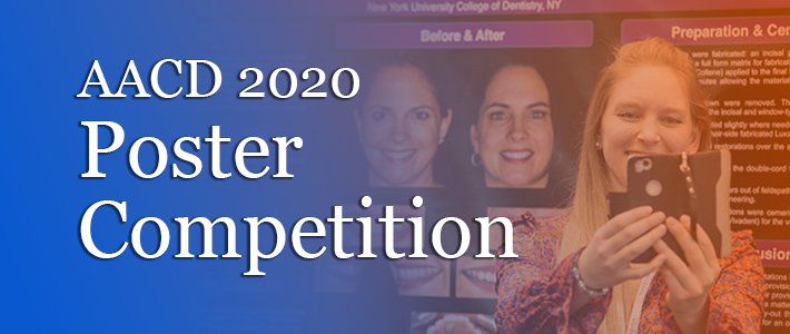
The American Academy of Cosmetic Dentistry (AACD) is proud to recognize the hard work and dedication of the following students for their work on research that was submitted for review in competitions that were to be held at the 36th Annual AACD Scientific Session in Orlando, Florida. The AACD Poster Competition creates an outlet for undergraduate and postgraduate students, junior dental faculty, along with clinicians and researchers, to share their scientific findings and to promote greater interaction within the dental community. Even though we were not able to meet in person, we still wanted to share this exciting research with everyone.
Clinical Cases
Restoring a Smile: Facial Integration Design
 Nicolas Aguilera, DDS | Post-Graduate | University of Rochester
Abstract: Successful comprehensive cosmetic dentistry relies on meticulous diagnosis, carefully sequenced treatment planning and the highest level of treatment execution. Many conditions need to be addressed prior to designing a new smile. The implementation of Digital Smile Design can strength diagnostic vision, improve communication, and enhance predictability. Pre-visualization is particularly indicated in cases where modification of the anterior teeth is required.
Faculty Mentors: Alejandro Sanchez-Lara, DDS and Hans Malmstrom, DDS
Layering Composite for Ultimate Aesthetics in Direct Restorations
Hanan Alalawi, BDS | Post-Graduate | Nova Southeastern University
Abstract: Direct composite veneers have gained an important role in clinical applications due to its excellent aesthetics. It allows dentists to add to tooth surfaces either to close gaps or reshape the tooth without cavity preparation. In this case, 13-year-old female presented with space between laterals and canines on both sides. Wax-up was done for #7 and #10 and palatal index was created. 38% phosphoric acid and Optibond FL primer and bonding agent were used. Vit-l-escence composite system shade (Dentin A2 + Enamel PA) was placed using layering technique to enhance the anesthetic outcome.
Faculty Mentor: Cesar Gonzalez, DDS
Crown Therapy, an Alternative to Restore Amelogenesis Imperfecta
 Maria Claudia Alvarado, DDS | Post-Graduate | University of Connecticut
Abstract: A 21-year-old female presented to the University of Connecticut, School of Dental Medicine seeking to improve the color of her teeth and repair the fractures existing on both arches. Intra and extra oral examinations and an esthetic evaluation were performed. The patient was diagnosed with Amelogenesis Imperfecta. Direct bonding and whitening were ruled out as effective solutions. A treatment plan was developed which addressed the esthetic issues and diagnosis while respecting function and biology. Ultimately, the patient was treated with a combination of Lithium Disilicate and Zirconia crowns.
Faculty Mentor: Sergio Sanches, DDS
Esthetic Restorations Using Lithium Disilicate Crowns on Discolored Teeth
Mariannina Savoca Astudillo | Undergraduate | Nova Southeastern University
Abstract: 35 years old female patient who presented in Nova Southeastern University dental clinic with the chief complain my two front teeth have changed color. Patient stated she received physical trauma on the mouth during an altercation when she was younger. After the incident, she noticed that teeth #9 and #10 were getting darker over time. As a consequence, both teeth were necrotic and #9 had a periapical pathology. Patient is healthy, ASA I. Takes multivitamins once a day. She is allergic to cat and denies any alcohol, smoking or drug use. After finished this case I was able to achieve good esthetic and function with two lithium disilicate crowns.
Faculty Mentors: Tulia M. Gonzalez, DDS and Dr. Michael Patten, DDS
Enhancing Tooth Shape and Shade Using Feldspathic Porcelain
 Vincent Avallone | Undergraduate | New York University College of Dentistry
Abstract: When evaluating a smile, it is important not only to look at the teeth themselves, but to look at the patient as a whole. This patient presented with small, spaced teeth, lip augmentation with Juvéderm (hyaluronic acid), muscle guarding from years of hiding her smile, and a very light skin complexion. By taking these extra factors into account, we are able to enhance the patients overall aesthetic at a macro level by redesigning the size, shape, and shade of her smile that fit her best. Patients Chief Complaint: I dont like my smile or the spaces behind my front teeth. After a thorough evaluation and treatment planning, our treatment goal was to enhance the overall shape and shade of the smile while closing the spacing seen in the anterior dentition. Using Feldspathic Porcelain, 9 veneers and 1 full coverage restoration were designed in order to achieve these aesthetic treatment goals while maintaining proper physiologic functionality.
Faculty Mentors: Kenneth Magid, DDS, FICD and John Calamia, DMD
Esthetic Rehabilitation of Maxillary Anterior Teeth Using a Minimal Invasive Approach: Ceramic Veneers and a Ceramic Crown
Behshid Bahraini, DDS | Post-Graduate | Tufts University School of Dental Medicin e
e
Abstract: This case report describes a minimally invasive approach when treating a 57 year old female patient with esthetic concerns about her maxillary anterior teeth shape, size and color. A comprehensive exam, facial and smile analysis, diagnostic wax up and mock up were completed. The treatment plan consisted of in-office and take home tooth whitening treatment, followed by feldspathic veneers on teeth #6, #7, #8, #9, #10, #11, an IPS E-max lithium disilicate crown on tooth #12 and a direct composite restoration on tooth #5 ensuring a conservative approach while achieving high esthetics results.
Faculty Mentors: Aikaterini Papathanasiou, DMD, DDS and Panagiotis Papaspyridakos, DDS, MS, PhD
Aesthetic Management of Proclined Anterior Teeth Utilizing Zirconia-Ceramic Fixed Bridge
 Dina Banakhar, BDS | Post-Graduate | Nova Southeastern University
Abstract: Clinical Case Presentation: A 43-year-old female patient who was very much dissatisfied with her teeth appearance presented with a chief complaint of My gums are receding and I want to replace my Maryland bridges in the front. On clinical examination, it was found that there was a generalized proclination of anterior teeth and recession. Radiographic examination showed tipping of the anterior roots due to previous orthodontic treatment. Case was managed by an anterior connective tissue graft, in-office bleaching, and to replace #6-8 & #9-11 Maryland bridges with Zirconia-Ceramic fixed bridge #6-11. Final bridge was cemented using (GC Fuji PLUSï).
Outcome: This multi-disciplinary approach had a positive impact on patients smile and it ultimately enhanced her aesthetic and self-confidence.
Faculty Mentor: Aryia Amini, DMD
Implant Rehabilitation in the Esthetic Zone: Missing Canines in the Anterior Maxilla
 Ayman Banjar, BDS | Post-Graduate | Tufts University School of Dental Medicine
Abstract: A 29-year-old female patient presented for rehabilitation of her congenitally missing maxillary canines referred by the Orthodontic Department. After comprehensive diagnostic work-up, the placement of two implants was planned after managing the spaces with orthodontics. After 2nd stage surgery, soft tissue conditioning was done with provisional crowns on implants #6 and 11 for a period of 3 months. Definitive rehabilitation was completed with combination of with veneers and single implant crowns. Lithium disilicate was the material of choice for the veneers and monolithic zirconia for the implant crowns.
Faculty Mentors: Aikaterini Papathanasiou, DMD, DDS and Panagiotis Papaspyridakos, DDS, MS, PhD
Camouflaging Congenitally Missing Lateral Incisors with Porcelain Veneers
 Parsa Basseri | Undergraduate | New York University College of Dentistry
Abstract: The contemporary abilities that dental professionals have in augmenting smile appearance introduce new benchmarks in cosmetic dentistry. As a tooth can be transformed into another form by restorative efforts, special care must be taken to ensure adequate preparation, gingival harmony and orthodontic stability when designing the illusion of aesthetic dentition. With digital photographic planning, active patient engagement, meticulous dental preparation and detailed laboratory communication, porcelain veneers can achieve the functional and aesthetic demands of patients who may have congenitally missing teeth.
Faculty Mentor: John Calamia, DMD
Honorable Esthetic Achievement Poster Student
The Art and Science of Matching Anterior Teeth Using Layered Zirconia Restorations
Yasko A. Darkoue, BDS, MS | Junior Faculty | University of Missouri 
Abstract: Almost 60 years ago, Weinstein invented metal ceramic restorations which was a breakthrough in esthetic restorative dentistry. Since then, dental restorative materials evolved, technology and techniques were also improved. Nowadays, Layered Zirconia Restorations offer an inherent strength while esthetics can also be optimized to tackle the challenge of matching natural teeth. This poster will discuss the art and science used to match central and lateral incisors of mild tetracycline stained case. The poster will also discuss how to tackle chipping of porcelain, demystifying bonding to zirconia, and a novel shade communication technique using grey reference cards, polarizers and digital photography.
Faculty Mentor: Celin Arce Urena, DDS
Esthetic Smile Improvement Utilizing Crown Lengthening and Porcelain Veneers
 Stacey Davidyants | Post-Graduate | New York University College of Dentistry
Abstract: A 30-year-old female patient in good health presented to NY Presbyterian Brooklyn Methodist Hospital seeking a change in her smile. One year prior, she was struck by a vehicle as a pedestrian, causing class II Ellis fractures to teeth #8 and #9. She was interested in a treatment option that incorporated her entire smile. Treatment goals included correction of midline cant, adjustment of occlusal cant, reduction of gummy smile, closure of diastemas, and improvement upon color, shape, and texture of the teeth. Treatment was completed with periodontal crown lengthening on teeth #4-13, followed by IPS e.max veneers on teeth #4-13.
Faculty Mentor: Kristine Hassan, DDS and Joseph Izzo, DDS
Minimally Invasive Approach for Treatment of Dental Fluorosis Using Tooth Whitening, Microabrasion and Porcelain Veneers
 Adnan Hakim | Post-Graduate | Tufts University School of Dental Medicine
Abstract: Dental fluorosis often presents with a combination of white and brown stains. Depending on the depth of the lesion, different treatment protocols can be implemented to improve the appearance of the teeth. This case report describes a minimally invasive approach on a 36 year old female patient with esthetic concerns regarding the shape and fluorosis discolorations of her maxillary anterior teeth. A comprehensive exam, smile analysis and diagnostic wax up were completed after which a minimal invasive treatment plan was conducted. It consisted of tooth whitening followed by microabrasion of teeth #6 and #11 and feldspathic porcelain veneers on teeth #7,8,9,10. Optimum esthetic results and high patient satisfaction were achieved with this conservative approach.
Faculty Mentors: Aikaterini Papathanasiou, DMD, DDS and Aikaterini Kostagianni, DMD, DDS, MS
Masking Dark Stump Shades with Translucent Zirconia
 Dorothy D. Hino | Undergraduate | University of Texas School of Dentistry
Abstract: It is not unusual for a patient's chief complaint to be esthetic in nature. Many variables need to be taken into consideration when addressing a patient's esthetic complaint. The clinician needs to consider whether the patients desires are clinically ethical and possible to achieve. The objective of this case is to demonstrate how to create an esthetic smile with a cohesive restorative shade using zirconia and porcelain to mask out dark stump shades. In this case, the communication with the dental lab and the patient played an important role in the successful outcome.
Faculty Mentor: Joe C. Ontiveros DDS, MS
Creating Function and Esthetic Provisional Restorations for Predictable Smile Enhancement Outcome
 Manar Tariq Karawi, BDS | Post-Graduate | Nova Southeastern University
Abstract: The presented case depicts defective PFM crowns #8, 9 of a 55-year-old-female presented with bone and soft tissue deficiency. Central incisors were extracted and replaced with implants and Guided Bone Regeneration, followed by immediate temporization. Lithium-Disilicate (IPS-e.max) screw-retained implant crown with Ti-base Zirconia custom abutment were used for #8,9. Porcelain veneers #7,10 were prepared to compensate space discrepancy between lateral and central incisors. Veneers were fabricated with Lithium-Disilicate (IPS-e.max) then bonded with Variolink Esthetic LC System.
Faculty Mentor: Aryia Amini, DMD
Esthetic Rehabilitation of a Smile with Multiple Implant Crowns and Ceramic Restorations
 Ahmed Haney Katib | Post-Graduate | Nova Southeastern University
Abstract: The presented case depicts a challenging esthetic issue. 61-Year-old male with implant abutments for #7,10, old defective porcelain crowns on #8,9 and defective class V resin restorations on #6,11 with gingival recession in the area. A mockup was done for the six anteriors. Preparation and temporization were done for teeth #6-11. #6,11 IPS emax Veneers, #7,10 IPS emax implant crowns with zirconia abutments & #8,9 IPS emax full crowns. Due to patient financial limitations, only six anteriors were restored for the time being and a temporary partial denture was fabricated while he undergoes implant placement of posteriors. Outcome: Multidisciplinary treatment aimed to restore esthetic and function. Establishing a good lab communication with photos and models to achieve best esthetic results.
Faculty Mentor: Aryia Amini, DMD
Restoration of Peg Lateral Incisors with Asymmetrical Spacing
Riley J. Robinson, DDS | Post-Graduate | University of Utah
Abstract: A 26 year-old female, presented with staining/fracturing of her peg lateral incisor composite build-ups and a chief complaint of, I want my front teeth fixed. A treatment plan was developed to replace the unaesthetic/defective restorations with EsthetX composite veneers to restore asymmetrical lateral incisor spaces. A wax-up was completed, and a lingual stent was fabricated to be used as a template for the final restorations. The definitive veneers were designed and fabricated to satisfy the patients chief complaint while maintaining a natural appearance. The patient was pleased with the improvement in overall tooth shape, color, and time efficiency.
Faculty Mentor: Craig Proctor, DDS
Breaking Bridges, Implanting Smiles: Functional and Aesthetic Rehabilitation After Maxillary Anterior Trauma
 Ryan Lisann | Undergraduate | Harvard School of Dental Medicine
Abstract: 50 y/o male presented with 8-x-10 FPD in hand: My bridge broke and my tooth is sensitive. Can you help me? The crown preparation for #10 fractured millimeters from the gingival margin and #8 was intact. 8-x-10 FPD was poorly shade matched with existing PFM crown on #7. An implant was placed at site of #9, and RCT with cast post and core completed on #10. After implant osseointegration, #7-10 were temporized individually with temporary on #9 to develop emergence profile. 4 PFM crowns were delivered with screw-retained abutment on #9. Collaboration involved prosthodontic, endodontic, periodontic, and implantology departments.
Faculty Mentors: Peter Grieco, DDS and Armando Pardo, DMD, & Hiroe Ohyama, DDS, DMD, MMSc, PhD
Restoring Fluorosis-Stained Teeth with Porcelain Veneers
Gili Litwack | Undergraduate | New York University College of Dental Medicine
Abstract: Chief Complaint: I am so embarrassed to smile because of the color of my teeth. Dental Fluorosis manifests itself by too much ingestion of fluoride resulting in disturbances in enamel mineralization. During developmental stages of tooth formation, high fluoride intake creates intrinsic yellow to dark brown staining of ones external enamel, causing a rather unaesthetic appearance. Fluorosis was suspected to be the cause of discoloration due to high fluoride concentration in the water (3.0-6.5 mg/L - far higher than the World Health Organization recommended 0.5-1.5mg/L) in the area in Yemen where the patient grew up. This suspicion was confirmed via slight abrasion of the enamel, easily removing some staining. The amount of enamel reduction necessary to completely remove the stains - both facially and proximally - required subsequent placement of a dental restoration.
Faculty Mentors: John Calamia, DMD, Fred Puccio, DDS, Kenneth Magid, DDS, and Larry Passaro
Veneers from #7-10 to Manage Anterior Open Bite
 Rafael Martinez | Undergraduate | Nova Southeastern University
Abstract: Patient with an anterior open bite and a chief complaint: "I don't want a space between my upper and lower front teeth". Lithium Disilicate veneers from #7 to #10 were planned to close the space and eliminate the inverse smile. Diagnostic wax up was done making sure that the future veneers were out of contact during excursive movements, and the length of the centrals were determined using the golden proportion based on the central width. Preparations were scanned using CAD/CAM and cemented using Variolink. Patient was really satisfied with outcome and is planning to get lower veneers.
Faculty Mentor: Jorge Rodriguez. DMD
Anterior All Ceramic Crowns: Smile Design Rules
Chintan Patel | Undergraduate | University of the Pacific
Abstract: Diagnosis and proper treatment planning is the key to success in any esthetic restorative case. This case involves full mouth rehabilitation with high esthetic demands from the patient. It is important to take an organized systemic approach to identify and resolve any functional and esthetic problems. Our goal for the esthetic zone restorations is not only managing the disease but also to achieve the best functional and esthetic outcome.
Faculty Mentor: Kalid Aziz, DDS, MS
Achieving Aesthetic Excellence with PFZ
 Andrew Powell | Undergraduate | Nova Southeastern University
Abstract: This case report details some of the challenges dealing with necrotic stained teeth in the aesthetic zone. This case was managed using natural opaquing properties of porcelain fused to zirconia crowns.
Faculty Mentors: Randy Lichtman, DDS, Diego Dalla Bona, DDS, PhD, and Steven Milhauser, DDS
Honorable Esthetic Achievement Poster Student
Establishing Aesthetic and Functional Harmony with Lithium Disilicate Restorations
Laura C. Rhein | Undergraduate | New York University College of Dentistry
Abstract: Utilizing a systematic approach to smile evaluation and dentofacial analysis is essential in the treatment planning stages of esthetic treatment. It is important to attentively listen to patients share their dissatisfactions with their current smiles as well as their goals and expectations for prospective treatment.
Patients chief complaint: My front veneer broke and I need to get a new one made.
Using questionnaire-style questions, the patient could share additional complaints about her smile, including her frustrations with years of ineffective bleaching treatments and the varying colors in her smile. Further examination revealed generalized attrition resulting in a loss of canine guidance. In this patients case, we aimed to establish esthetic and functional harmony with lithium disilicate restorations.
Faculty Mentor: John Calamia, DMD, Nicholas Giannuzzi, DDS and Kenneth Magid, DDS, FICD
Indirect Esthetic Restorations; Anterior Zirconia Crowns
 Kavita Sharma | Undergraduate | Columbia College of Dental Medicine
Abstract: A 52 year old female patient presented with a chief complaint of "diastema between her maxillary central incisors, two fractured all ceramic crowns on teeth #7 and #8 and dissatisfaction with the color of her anterior maxillary teeth". An esthetic analysis, diagnostic wax up and mock up was done as a part of the treatment planning for the case. Treatment included, surgical crown lengthening of her anterior teeth followed by restoration of teeth #6 -11 with Monolythic Zirconia crowns (Katana, UTML). The treatment successfully closed the diastema, accomplished the desired esthetic results and met the patient's satisfaction.
Faculty Mentor: Mark L. Pitel, DMD, FAGD, FACD, FADFE
Digital Wax Up Using 3D Sculpting Based Computer Assisted Design
Yousof Sinjab | Undergraduate | Nova Southeastern University
Abstract: Dental mockups play a valuable role in modern dentistry, with applications in both esthetic and prosthetic treatment. The mockup allows the dentist to evaluate esthetics, function and speech before the delivery of a definitive restoration. Whether it is used for a simple crown, implant, or even a complete smile makeover, mockups help to manage patient expectation and facilitate between patient, dentist, and technician. communication.
Faculty Mentor: Steven A. Milhauser, DDS
Conservative Management of Diastema and Tetracycline Staining in Aesthetic Zone
 Cuong To | Undergraduate | Virginia Commonwealth University
Abstract: Patient presented with diastema, tetracycline staining, and failing composite restorations. Clinical evaluation revealed inadequate tooth form and lip support. Six lithium disilicate veneers (#6 to #11) were recommended to correct functional and aesthetic desire. Mock-ups using bis-acrylic resin from conventional and digital diagnostic wax-ups were used to assess occlusion, aesthetic, and phonetics. Teeth were then prepared and intraorally scanned with Planmeca. Six lithium disilicate veneers were designed, tried in, and bonded using VarioLink LC system. Patient was pleased with the functional and aesthetic outcome, especially the improved lip support that provided a younger appearance.
Faculty Mentors: Sompop Bencharit, DDS, MS, PhD and Rami Ammoun, DDS, MS
Smiling Again: Replacing 25 Year Old Gold Crowns with Esthetic Ceramics
Natalie Vos | Undergraduate | University of Texas School of Dentistry
Abstract: Patient presented with gold crowns on his maxillary front teeth and wants to replace them with natural looking teeth. Original crowns were placed about twenty-five years ago by a non-clinician. Our treatment plan consisted of assessing the patients chief complaint along with evaluating esthetic and functional demands to better improve his overall oral health. The patient understood the risks involved with removing the current gold crowns. This current case presents the transformation of replacing 25 years of gold with anterior ceramic restorations.
Faculty Mentor: Joe Ontiveros DDS, MS
Honorable Esthetic Achievement Poster Student
Transforming Canine to Canine: Replacing Broken FPD While Replacing Three Class V Lesions with Veneers
 Alan M. Williams | Undergraduate | The University of Texas School of Dentistry at Houston
Abstract: 58 year old male presents with chief complaint of I want veneers and to have my crooked bridge fixed. Patient had previous bridge fall out and superglued it back on himself. Previous Class V composite restorations on #6-#8 have undergone marginal staining. Patient desired to have bridge straightened out and #6-#8 veneers to match the color of the bridge. Finances were not a factor. Existing bridge was removed to evaluate condition of #9 & #11, both deemed restorable. Lithium disilicate veneers #6-#8 and FPD #9-11 with basic shade 1M2 and incisal 1M1 were fabricated. Occlusal guard was encouraged after treatment.
Faculty Mentor: Dr. Joe Ontiveros DDS, MS
Prosthetically-Driven Implant Placement and Restoration of the Esthetic Zone
Matthew Yeung, DDS| Post-Graduate | Dental College of Georgia
Abstract: A 46 year-old Hispanic male presented to clinic as a workers compensation case, missing teeth # 7-9 due to trauma. Under the circumstances, treatment was limited to the affected area and there was a time constraint due to an expiring VISA. An in-house digital workflow was followed to plan, design and print a surgical guide for implants at site # 7 and 9. A fully guided surgical protocol was followed, and custom healing abutments were fabricated to sculpt the gingival tissues. After 12 weeks from implant placement, a screw-retained, layered zirconia FDP was delivered that satisfied phonetics, occlusion and esthetics.
Faculty Mentor: Mario Romero, DDS
Materials Science Cases
Flexural Strength of Three-Unit Monolithic Zirconia FPD with Different Size of Connectors Sintered in SpeedFire Oven
.JPG) Behshid Bahraini, DDS | Post-Graduate | Tufts University School of Dental Medicine
Behshid Bahraini, DDS | Post-Graduate | Tufts University School of Dental Medicine
Abstract: The aim of this in vitro study was to investigate the flexural strength of three-unit monolithic zirconia FPD with different size of connectors sintered in SpeedFire oven.
Materials & Methods: Thirty-six, three-unit zirconia (In-Coris TZI C Medi S A1, Sirona Dental Systems GmbH, Bensheim, Germany) FPDs with different connector sizes were milled, sintered, and glazed. Three groups were formed based on the connector size: Group A: 3x3 mm, Group B: 3x4 mm and Group C: 4x4 mm with regard to connector thickness. Flexural strength was calculated in MPa with the aid of a universal testing machine (Model 5566; Instrom Corp, Canton, MA) along with the determination of mode of failure. The force was applied at the center of the bridge at a crosshead speed of 0.5mm/min, until it cracked.
Results: Group A had the highest mean flexural strength (2929.96 MPa with a Standard deviation of 355.17). Welch's ANOVA result was not statistically significant (p= 0.053). 74.3% of the fractures occurred in the connector part of FPDs and 25.7% happened in the margin of restorations. Concerning the mode of failure, based on Fisher's exact test, the difference between groups was not statistically significant. (p=0.717).
Conclusions:
1- No statistically significant difference was observed between the flexural strength of three-unit FPDs with different connector sizes sintered in the SpeedFire oven.
2- All of the 3-unit zirconia FPDs with connector sizes of 3x3 mm, 3x4 mm, and 4x4 mm, which were sintered with the short sintering protocol, were clinically acceptable in terms of flexural strength.
Faculty Mentors: Aikaterini Papathanasiou, DMD, DDS, Aikaterini Kostagianni, DMD, DDS, MS, Matthew D. Finkelman, PhD and Ala Ali, BDS, MSc, DSc, DMD
Accuracy of 3D Printed Casts from Digital Implant Scans versus Stone Casts from Conventional Impressions: A Comparative Study in the Anterior Maxilla
Ayman Banjar, BDS | Post-Graduate | Tufts University School of Dental Medicine
Abstract: This is an In-vitro study comparing the accuracy of 3D printed casts to conventional stone casts. Maxillary master cast was fabricated with two implants (Regular CrossFit®, Straumann). Conventional impressions were taken to fabricate stone casts. Trios3shape intraoral scanner was utilized to take digital impressions for printing casts using Form2 and Varseo S 3D printers. All casts were digitized with a reference scanner, and the STL files were superimposed to the master cast dataset in an inspection software to assess the 3D deviation. The conventional group had the lowest mean value followed by Varseo S group. The Form2 group had the highest mean value. Post-hoc test revealed significant difference between Varseo S and Form 2 groups.
Faculty Mentors: Yo-Wei Chen, DDS, MSc, Aikaterini Kostagianni, DDS, DMD, MS, Matthew D. Finkelman, PhD, Aikaterini Papathanasiou, DMD, DDS and Panagiotis Papaspyridakos, DDS, MS, PhD
Evaluation of Flexural Strength of Zirconia-Reinforced Lithium Silicate Glass Ceramic and Lithium Disilicate Glass Ceramic Restorations Fabricated with Heat Pressing Technique
 Adnan Hakim | Post-Graduate | Tufts University School of Dental Medicine
Abstract: This in-vitro study was conducted to compare the flexural strength of zirconia-reinforced lithium silicate ceramic (Celta® press, Dentsply, Germany-Group 1) with lithium disilicate ceramic (IPS e.max Press, Ivoclar-Vivadent, Liechtenstein-Group 2) and to investigate the influence of different surface treatments on the fracture resistance. Specimens were fabricated with pressing technique and randomly divided into four subgroups for each group: No treatment (A), Glazed (B), Polished(C), Polished and Glazed (D). Biaxial flexural strength test was performed and results were calculated in MPa. IPS e.max showed statistically significant higher flexural strength than Celtra press (p < 0.001). For surface treatment, the difference between the groups was statistically significant (p = 0.007) with polished and glazed group showing the highest mean value.
Faculty Mentors: Aikaterini Kostagianni, DMD, DDS, MS, Matthew D. Finkelman, PhD, Aikaterini Papathanasiou, DMD, DDS, and Ala Ali, BDS, MSc, DSc, DMD
Shear Bond Strength Evaluation of Contemporary Dental Cements on Zirconia
 Thuong Huynh | Undergraduate | University of Tennessee Health Science Center College of Dentistry
Abstract: Purpose of this study will be to evaluate the shear strength bonding on monolithic Zirconia utilizing six different dental cements.
Methods: Thirty rectangular (8x12x2mm) Zirconia specimens per material: BruxZir, Katana, ZirCAD, and Lava were mounted in acrylic (SamplKwick) and abraded with 50μm alumina particles for 10 seconds, then air dried. Each specimen was prepared with a bonding agent (MonoBond Plus) and subsequent cement (Multilink, BioCem, GCem, SpeedCEM, RelyX, and Panavia) formed into plastic cylinder and oriented for shear bond testing. Cements were tact-cured for 20 seconds with an Elipar DeepCure-S curing light. After the first cure, the plastic cylinder was removed, and the cement was cured again from each direction for 20 seconds. The bonded zirconia specimens were stored in a solution of deionized water at 37°C for 24 hours to allow the sample to cure. Specimen bonded with GCem was stored in a separate container because of the glass ionomer base of the resin. Shear bond strength testing was performed on an UltraTester Bond Strength Testing Machine with a speed of 1.0mm/minute. Maximum stress (MPa) data were compared and statistically analyzed using a two-way repeated measure ANOVA SNK with P<0.05 for significance.
Conclusions: Significant differences in the mean of the shear bond of the tested materials were observed. Within the testing conditions of this study, Zirconia bonding with an array of different cements only demonstrated a significantly higher difference in shear bond strength for GCem paired with Katana. When using BioCem, a significant lower shear bond strength was observed with BruxZir (P=0.02), Katana (P<0.001), and Zircad (P=0.034). Significant higher shear bond strength for GCem paired with Katana than other tested materials.
Faculty Mentor: Mohamed A. Shafter, DDS
Efficacy of Remineralizing Agents on Bovine Enamel Exposed to High Cariogenic Challenge: In Vitro Study
 Shaan Sehgal | Undergraduate | Harvard School of Dental Medicine
Abstract: Dental caries is one of the most prevalent chronic diseases worldwide involving the dynamic process of demineralization and remineralization. This study evaluated the remineralizing potential of five treatments under constant cariogenic challenge. It is hypothesized that fluoride products have a greater remineralizing potential than casein-derived ones. Fifty-five bovine fragments were immersed in an acetate buffer (pH 4.0) and randomly divided into five groups (n=11): G1=control, G2=Duraphat (Colgate), G3=1.23% Acidulated phosphate fluoride (Flugel,DFL), G4=MI Paste® Mousse (GC), G5=MI Paste® Plus (GC). Treatments were applied 3x over a period of 21-days and pH cycling was performed in-between. Samples were evaluated through optical microscopy, Fourier transform infrared spectroscopy, and with a microhardness tester. Data was compared by calculating hardness variation, a Holm-Sidak test, and descriptive analysis. It was found that Flugel was the only treatment effective in remineralizing lesions supporting literature that shows fluoride products can reverse early lesions.
Faculty Mentors: Hiroe Ohyama, DDS, DMD, MMSc, PhD and Shelyn Akari Yamakami, DDS, MSc